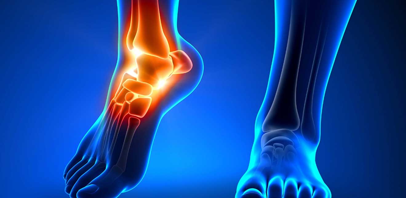
Deformities
Talipes valgus
It is a condition in which a farther segment of the bone is angulated outwards from the joint away from the midline of the body. Talus valgus is usually a secondary abnormality; it is mostly due to as a compensation of primary abnormality in the lower limb. Few primary conditions are Genu valgus(Knocked knees), genetics The treatment of this condition involves eliminating the need of compensation due to the primary condition. In addition to primary condition treatment, a specially designed orthotic have to be used to accommodate the deformity.
Talipes Varus
It is the condition in which the farther segment of the bone is angulated inwards from the joint towards the midline of the body. A notable type of talus varus is clubbed foot medically called as clubbed talipes equinovarus (CTEV)—it is a congenital deformity found in the newborns, where one or both feet are rotated inwards. It is a mix of bones and soft tissue deformity, which makes it a complex condition. If not managed or treated successfully, it can become a severe lifelong permanent impairment. If left uncorrected, as the child grows, he /she will start putting the weight on the sides or front of feet making those areas hard and painful.
The management of this condition can start as early as it is detected (early infancy). There are two options of treatment:
- The conservative management: Once the CTEV is successfully diagnosed, the surgeon starts with specialized orthotic shoes, where the shoes keep the feet in outward rotated position. Another type of method is using the cast application. The cast application used in a series of application where it is changed every week with every week has different style of cast and series of manipulations and the position of the feet changes with every cast. Finally, a small tenotomy (a small cut is made on a tendon) of Achilles tendon is done. To have better results with this method, it is mandatory to have a close follow up for at least 4-5 years to maintain the achieved. Since, there are 50% chances of the feet to return to the deformed state.
- Surgical intervention: Surgery is opted by individuals who wish to have direct results and also if, non operative treatment fails or achieve incomplete correction of deformity. There are multiple procedure can be applied to correct the deformity. Your surgeon depending on the required correction decides the exact procedure and extent of correction.
Dr. Zia has been successfully treating foot deformities with utmost care and precise choice treatments. He is a firm believer in eliminating deformities by appropriate treatment and careful planning.
Ligament Injuries of the foot
There are more than hundred muscles, ligaments and tendons in a foot. In harmony with the bones, the ligaments, muscles and tendons all of which work together to provide support, mobility and balance. The main ligaments of the foot are:
- The plantar fascia— it is the longest ligament of the foot that runs from the toe to the heel. It forms the arch of the foot that contracts and stretches to help us balance and provides strength while walking or running.
- Plantar calcaneonavicular ligament— It is the ligament of sole which connects the calcaneus(heel bone) , navicularis and head of talus(bone under the ankle).
- Calcanecuboid ligament—it is the ligament which provides support to the plantar fascia to maintain the arch.
- Lisfranc ligament— It is an interosseous ligament that runs from cuneiform (one of the tarsal bone) to the base of second metatarsal. It has a very crucial role in foot anatomy; Lisfranc ligament stabilizes the second metatarsal and help in maintenance of the foot arch.
Plantar fasciitis
It is the inflammation of plantar fascia (the ligament that runs along the sole from toe to heel). It happens due to chronic overuse leading to micro tears at the origin of the fascia, repetitive trauma leading to recurrent inflammation.
Symptom of plantar fasciitis is the sharp heel pain, especially after waking up and at the end of the day due to prolonged standing. It can be in one foot or in both feet. A through of physical examination of the foot and ankle can help in diagnosis additional to X-rays and MRI these can help in planning surgery if required.
The treatment of plantar fasciitis is rather conservative than surgical. Surgery is required in more extreme cases. The non-operative treatment involves:
- Physical therapy, A proper stretching program
- Non-steroidal pain killers
- Steroid injection in the affected area.
- Foot orthrosis- these are pre-fabricated shoe insert helps in cushioning he heel and maintaining the arch.
Lisfranc Injuries
Lisfranc ligament with the tarsals and metatarsals forms a joint complex consist to three articulations. It gets easily injured during motor vehicle accident involving the foot, fall from heights, and injuries while playing running sports.
Symptoms of Lisfranc injuries are typical severe pain, inability to bear weight. A through physical examination with motion and instability tests can help in diagnosis. An X-ray of the foot can clearly aid in diagnosis with its typical appearances and signs. Further examinations like CT and MRI can help in preoperative planning.
The treatment of the injury depends on the extent of it. A non-operative management can be done in cases with no displacements on weight bearing and in cases where surgery is contraindicated. The non-operative treatment will be of splinting and cast immobilization for 4 weeks – 8 weeks.
The surgical treatment can be done with open reduction and internal fixation.
In this procedure, the surgeon cuts open the skin and exposes the fracture carefully. The bone fragments and injured/damaged ligaments are repositioned and normally realigned in their natural position.
Surgical fixation may be fixed with many devices: Pins, screws, plates, rods and external fixators. And in few cases the bone gets severely damaged and that may create a gap between two bony pieces. In these cases, a bone graft is added to the fracture reduction to help the repair process.
Diabetic foot
A diabetic foot is a complication of diabetes mellitus (type 2 diabetes). It is presents with multiple pathologies; uncontrolled/poorly controlled blood glucose levels, peripheral neuropathy, peripheral artery disease, and infection.
Due to peripheral neuropathy, patients have reduced sensation to pain. This could lead to inability to feel pain from minor injuries and they remain undiscovered for a long while. In association with peripheral arterial disease, it could lead to poor circulation in the area and further worsen the health of the foot.
Diabetes has been noted as a poor immune state, where it is difficult for the body to fend off minor infections. Due to increase in pressure in the foot, a pressure ulcer can form and progress in spreading allover the foot. Foot infection has become most common cause of amputation in diabetes.
Prevention and treatment: Prevention of foot infection in diabetes involves optimistic control of blood glucose level with regular monitoring, feet hygiene and moderate exercise.
Treatment of diabetic foot is challenging and prolonged therapy. It may include specially designed orthopedic shoes, regular dressings of the wound, antibiotic therapy. Sometimes, in few cases there is a co-fungal infection, which requires antifungal treatment as well. Most diabetic foot infections need system antibiotic therapy with constant monitoring of infection and antibiotics to be used in proper dosage in order to avoid development of resistance.
Gouty Arthritis
It is a form of inflammatory arthritis; it develops in people with high uric acid levels. The higher level of uric acid in the blood stream get deposited in joints, in form of uric acid crystals. The deposition of crystals will trigger an inflammatory response in the joint, which gives rise to sudden sharp pain, tenderness, warmth and redness around the affected joint. The disease progress in to stages:
- Acute stage: It happens when there is sudden rise in uric acid level in the bloodstream, it happens mostly after binge alcohol consumption. The acute hyperuricemic stage makes the crystal deposition quite fast. The symptoms are; sudden sharp pain, tender and swollen joints. This condition is self-resolving — it may take up to 2 weeks. In here patient is advice to quit alcohol and restrict taking red meat diet and certain seafood.
- Interval Gout: It is a stage of gout where the inflammatory response is low. Although the pain and swelling might not be present in this stage. The low level inflammatory response is still damaging the joints. In future it might develop in to chronic gout. To prevent it from developing in to chronic gout, it is recommended to make few lifestyle changes and start medication.
- Chronic Gout: It is a long-standing condition where the deposition of uric acid crystals has been neglected for number of years. The gout attacks may occur frequently and the pain might not go away easily. Due to the long-standing condition—the joint damage may be severe, which can lead to loss of mobility.
Gout has been seen more in men than women. Few experts say it is due to the protective nature of estrogen. Other factors causing gout are: genetically inherited, poor lifestyle, high fructose diet and high red meat diet, gastric by pass surgery, obesity and alcohol consumption.
Diagnosis: diagnosis of gout is made by complete medical history and physical examination of the affected joint. Complete blood panels’ helps the doctor rule out other causes and check the uric acid levels in bloodstream. CT and MRI can help in assess the extent of joint damage and further treatment plan. Doctor might take out some joint fluid and examine it under microscope to confirm the presence of uric acid crystals; this method is the surest possible way to affirm the diagnosis of gout.
Treatment: Gout treatment requires twin disciplinary approach. One being the lifestyle modification and second is medication to ease the pain and decrease the uric acid level in the blood stream.
Lorem ipsum dolor sit ametcon sectetur adipisicing elit, sed doiusmod tempor incidi labore et dolore.
Lorem ipsum dolor sit ametcon sectetur adipisicing elit, sed doiusmod tempor incidi labore et dolore.
Lorem ipsum dolor sit ametcon sectetur adipisicing elit, sed doiusmod tempor incidi labore et dolore.


